REPRODUCTION GRADE 12 NOTES - LIFE SCIENCES STUDY GUIDES
Share via Whatsapp Join our WhatsApp Group Join our Telegram GroupREPRODUCTION
LIFE SCIENCES
STUDY GUIDES AND NOTES
GRADE 12
- Male reproductive system
- Activity 1
- Female reproductive system
- Activity 2
- Puberty
- Menstrual cycle
- Hormonal control of the menstrual cycle
- Activity 3
- Development of the foetus
- Activity 4
CHAPTER 4:REPRODUCTION
4.1 Male reproductive system
Figure 4.1 below shows the different parts of the male reproductive system and their functions.
Figure 4.1 Structure of the male reproductive system
Functions of testosterone
The testes produce the hormone testosterone, which has the following functions:
- Development of male secondary sexual characteristics, such as beard, pubic hair, deep voice and a muscular body.
- Stimulates the maturation of sperm cells.
Structure of a sperm cell
Figure 4.2 below shows the different parts of a sperm cell and their functions.

Figure 4.2 Structure of a sperm cell
Activity 1
Questions
- Name the accessory glands of the male reproductive system and give ONE function of each. (10)
- Name the organ where testosterone is produced. (1)
- Give TWO functions of testosterone. (2)
- Name all the parts of the sperm cell that are responsible for movement. State what the function of each part is. (4)
- Explain the role of the nucleus of the sperm cell in fertilisation. (3) [20]
Answers to activity 1
- Seminal vesicle ✔ produces a fluid that contains nutrients ✔ for the sperm cells, so that they have energy to swim.✔
Prostate gland ✔ produces an alkaline fluid ✔ that neutralises acids ✔ produced in the vagina, so that sperm cells are protected.✔
Cowper’s gland✔ produces mucus✔ that helps with the movement✔of sperm cells. (10) - Testes✔ (1)
- Testosterone is responsible for the development of male secondary sexual characteristics✔ and it stimulates the maturation of sperm cells.✔ (2)
- Mitochondria✔ provide energy for swimming.✔ Tail✔ moves in a whip-like fashion to propel the sperm cell forwards.✔ (4)
- The nucleus contains 23 chromosomes (n)✔, and fuses with the nucleus of an egg cell, which also contains 23 chromosomes (n)✔. The result is a zygote with
46 chromosomes (2n).✔ (3) [20]
4.2 Female reproductive system
Figure 4.3 below shows the different parts of the female reproductive system and their functions.
Figure 4.3 Structure of the female reproductive system
Activity 2
Questions
Provide the correct biological term for the following definitions.
- The inner lining of the uterus (1)
- Tube that connects the ovaries to the uterus (1)
- The structure that produces female hormones (1)
- The part where development of the embryo/foetus normally takes place in humans (1) [4]
Answers to activity 2
- Endometrium✔
- Fallopian tube✔
- Ovary/placenta✔
- Uterus✔ [4]
4.3 Puberty
Puberty is the period in humans in which they experience physical changes in their bodies in order to be capable of sexual reproduction.
Puberty in males | Puberty in females |
Stimulated by testosterone | Stimulated by oestrogen |
Growth of male sex organs | Growth of female sex organs |
Start of the production of sperm cells | Start of the menstrual cycle and production of ova |
Growth of pubic hair, facial hair and body hair | Growth of pubic hair |
Development of muscles and deepening of voice | Growth and development of breasts and widening of hips |
4.4 Menstrual cycle
The series of diagrams in Figure 4.4 below shows the events occurring in the ovary (ovarian cycle) and uterus (uterine cycle) during the menstrual cycle. The days are not exact, but are averages.
Figure 4.4 The menstrual cycle
4.5 Hormonal control of the menstrual cycle
The graph in Figure 4.5 below shows changes in the ovary, uterus and in the level of hormones during a 28-day menstrual cycle.
Figure 4.5 Hormonal regulation of the female reproductive cycle
The hormonal changes that take place at A, B, C and D in the graph in Figure 4.5 above are explained in Table 4.1 below.
| A | B | C | D | |
| Day 0-11 | Pituitary gland produces FSH which stimulates development of the follicle. | Follicle is developing to become a Graafian follicle containing an egg cell. | Oestrogen levels increase as the hormone is produced by the follicle. | Thickness of endometrium increases from day 7 (after menstruation has ended) as a result of oestrogen. |
| Day 11- 17 | FSH and LH (produced by the pituitary gland) levels are highest around day 14. | Follicle development is completed as a result of the influence of FSH by day 14. Ovulation is stimulated by high levels of FSH and LH on day 14. LH then stimulates the development of the corpus luteum. | Oestrogen levels reach a maximum towards day 14 until ovulation takes place, but then start to decrease because the Graafian follicle stops functioning. | Endometrium thickens further. |
| Day 17-28 | LH levels decrease and then remain constant to maintain the corpus luteum. | Corpus luteum produces progesterone. Corpus luteum gradually disintegrates since fertilisation does not take place. | Oestrogen levels increase again and then decrease towards the end of the cycle. Progesterone levels increase towards day 21. Progesterone levels decrease when corpus luteum disintegrates and stops functioning. | Progesterone prepares endometrium fully for pregnancy. Decreased progesterone levels from around day 21 cause endometrium to shed after day 28 by menstruation since no fertilisation took place. |
Table 4.1 Hormonal changes during the menstrual cycle
Activity 3
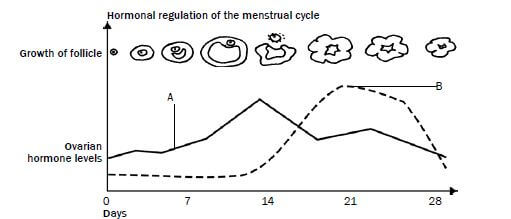
Study Figure 4.6 below and answer the questions that follow.
Figure 4.6 Hormonal changes during the menstrual cycle
- Name the hormones A and B. (2)
- Give reasons for your answers in question 1. (2)
- What event occurs on day 14? (1)
- Name the other two hormones involved in this cycle. (2)
- Did fertilisation occur during the cycle shown in Figure 4.6? (1)
- Explain your answer in question 5. (2) [10]
Answers to activity 3
- A - Oestrogen✔ B - Progesterone✔ (2)
- A: The Graafian follicle secretes oestrogen✔/Oestrogen reaches its maximum level before ovulation.✔
B: The corpus luteum produces progesterone✔/Progesterone reaches its maximum level after ovulation.✔ (2) - Ovulation✔ (1)
- LH✔ and FSH✔ (2)
- No✔ (1)
- Progesterone levels decrease✔ towards the end of the cycle.
The corpus luteum decreases✔ in size. (2) [10]
HINT:
Here is a hint to help you to remember the names of the two hormones:
- O stands for Oestrogen and when it is high, Ovulation occurs.
- P stands for Progesterone and when it remains high, there is a Pregnancy.
4.6 Development of the foetus
Figure 4.7 below shows the stages in the development of the foetus.
Figure 4.7 Stages in the development of the foetus
Explanation of Figure 4.7
- In the ovary a mature Graafian follicle bursts (usually on day 14 of the menstrual cycle) and releases an egg cell. This process is called ovulation.
- Fertilisation takes place high up in the fallopian tube. The egg cell (containing 23 chromosomes) and sperm cell (containing 23 chromosomes) fuse to form a zygote (containing 46 chromosomes).
- The zygote divides by mitosis to form a morula, then a blastocyst, and finally an embryo as it moves down the Fallopian tube.
- It takes about 5 to 7 days for the embryo to reach the uterus.
- In the uterus the embryo settles on the endometrium and sinks into it, embedding itself in the endometrium. This process is called implantation.
- After implantation, the embryo produces many finger-like structures called villi from the outer membrane of the embryo, which is known as the chorion.
- The villi grow into the tissue of the uterus to form a placenta.
- The placenta is attached to the embryo by the umbilical cord. It has 2 umbilical arteries (which carry deoxygenated blood from the embryo towards the placenta) and 1 umbilical vein (which carries oxygenated blood from the placenta to the embryo).
- The embryo is enclosed in a fluid-filled sac called the amnion. The fluid is called the amniotic fluid.
- After about 8 weeks, the embryo develops structures such as limbs and all the organs of the body. Now it is called a foetus.
- Gestation is the period between fertilisation and the birth of the baby. It usually lasts for a period of 9 months (39-40 weeks).
- The stages involved in the natural birth process are:
- Dilation of the cervix (labour)
- Expulsion of the foetus.
- Delivery of the afterbirth (placenta) and extra-embryonic membranes.
Activity 4
Questions
- On which day of the menstrual cycle does ovulation usually take place? (1)
- What happens to the Graafian follicle after ovulation? (1)
- Name the TWO hormones that are released by structures in the ovaries. (2)
- Give THREE functions of the amniotic fluid. (3)
- Give TWO substances that can move from the mother to the foetus through the placenta. (2)
- Give TWO substances that can move from the foetus to the mother through the placenta.
Answers to activity 4
- Day 14✔ (1)
- It changes into a corpus luteum.✔ (1)
- Oestrogen✔ and progesterone.✔ (2)
- The amniotic fluid protects the foetus against shock✔, drying out✔ and temperature changes.✔ (3)
- Oxygen✔, nutrients✔ (amino acids, glucose, other sugars), viruses✔ and drugs✔ (2)
- Carbon dioxide✔ and waste products✔ (urea). (2) [11]